DMAB-Nitrite for Tryptophan
The DMAB-nitrite histochemical method is a simple and very highly selective, almost specific, method for the amino acid, tryptophan. A blue product obtained with this technique is invariably accepted as proof that the blue stained material contains tryptophan.
DMAB is p-dimethylaminobenzaldehyde, also known as 4-dimethylaminobenzaldehyde, 4-Formyl-N,N-dimethylaniline and N,N-Dimethyl-4-formylaniline.
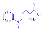
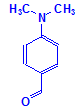
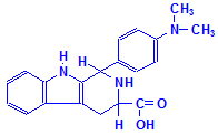
The leftmost structural formula above is of tryptophan, the central one is of DMAB, and the rightmost formula is of the initial reaction product that is produced by them. This reaction product is then oxidized with potassium nitrite to form an easily seen, blue colored compound of unspecified structure. It should be noted that formaldehyde and glyceraldehyde can also participate in similar reactions, so if this method is contemplated then formalin fixation should be kept short. The common overnight fixation in 10% formalin variants still permits the technique to give a positive result.
Materials
- p-Dimethylaminobenzaldehyde, 5% in concentrated hydrochloric acid
- Sodium nitrite (NaNO2), 1% in concentrated hydrochloric acid
Tissue Sample
15µ paraffin sections of tissues fixed up to 24 hours in one of:
- 1% trichloracetic acid in 80% ethanol
- 10% sulphosalicylic acid
- 10% neutral buffered formalin (6 hours preferred)
Protocol
- Bring sections to ethanol via xylene and ethanol and allow to just become dry.
- Place into DMAB solution for 1 minute
- Place in sodium nitrite solution for 1 minute.
- Rinse with tap water for 30 seconds.
- Dehydrate with ethanol.
- Clear with xylene and mount with a resinous medium.
Expected Results
- Tryptophan – blue
Notes
- Positive results should be seen in fibrin and fibrinoid, amyloid, Paneth cell granules, peptic cell granules, zymogen granules, muscle, neurokeratin and hair root sheath. Of these, only fibrin, including fibrinoid, and amyloid are extracellular materials which could be confused.
- When used to confirm that a material is amyloid, the positive material may be differentiated from fibrin by dye staining methods, i.e. a positive DMAB-nitrite stain with a congo red stain also positive would identify the stained material as amyloid with a high degree of certainty, as fibrin is not stained with that dye.
Safety Note
Prior to handling any chemical, consult the Safety Data Sheet (SDS) for proper handling and safety precautions.
References
- Adams, C.W.M., (1957),
A p-dimethylaminobenzaldehyde-nitrite method for the histochemical demonstration of tryptophane and related compounds.,
Journal of Clinical Pathology, v 10, page 56-62. - Pearse, A.G.E. (1968).
Histochemistry: Theoretical and Applied, Ed. 3, Volume 1.
Churchill Livingstone, London, England. - Davenport, H.A.. (1960).
Histological and Histochemical Technics,
W. B. Saunders, Philadelphia, USA.


